




August 11, 2025 -- When we set out to design the Micro-X Head CT, our goal was simple: to bring stroke diagnosis to patients faster, whether in an ambulance, a rural clinic, or a hospital emergency bay. But achieving this meant rethinking how CT scanners work at a fundamental level.

In conventional CT scanners, flat-panel detectors rotate around the patient on large gantries. These systems are incredibly effective but they’re also heavy, expensive, and mechanically complex. That makes them impractical for mobile environments where space, weight, and speed are critical.
To build a system compact and lightweight enough for stroke diagnosis on the move, we needed to do something never done before: create a curved detector.
Why Curved Matters
With a curved detector, we could achieve wide angular coverage using a fixed gantry.That meant we could eliminate the need for heavy rotating parts while still capturing high-quality projection images from multiple angles. It was a breakthrough in reducing size, weight, and power requirements, making the Head CT faster, lighter, and more portable.
But here’s the catch: there are no off-the-shelf curved X-ray detectors suitable for clinical CT imaging. Curved detectors have been explored in research or industrial uses (like inspecting pipelines), but not in medical imaging. So, we had to build one from scratch.
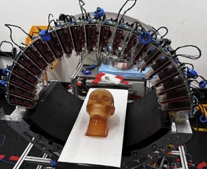 Head CT under development
In brief
Micro-X says its Head CT system, which is under development for stroke diagnosis, uses a curved detector to provide wide-angle image coverage with a fixed gantry rather than rotating mechanical components. The company co-developed a custom detector with FUJIFILM, using its flexible scintillator technology, and says a redesigned version weighs 72% less than the initial prototype. Micro-X also worked with Johns Hopkins University on reconstruction methods intended to address image artifacts caused by the detector’s curved geometry. The system combines the detector with Micro-X’s NEX technology to capture multiple projections rapidly without a rotating gantry. The company says that, to its knowledge, the Head CT is the first CT scanner designed for clinical use to feature a curved detector.
Key takeaways
- The Head CT is under development for mobile stroke diagnosis.
- Micro-X and FUJIFILM co-developed a custom curved detector using flexible scintillator technology.
- Micro-X says a redesigned detector weighs 72% less than its initial prototype.
- Johns Hopkins University worked with Micro-X on image reconstruction methods for curved geometry.
Partnering for Innovation
We partnered with FUJIFILM to co-develop a custom solution, leveraging their flexible scintillator substrate - the same material they use in their flat panel detectors. Our team worked closely with theirs to overlap two scintillators into a seamless curved array, paired with a dynamic readout system capable of capturing multiple exposures rapidly. This was crucial for integrating with our proprietary NEX technology and enabling fast reconstruction.
With further refinement, we engineered a new version of the detector using FUJIFILM’s technology, redesigned by Micro-X to be even smaller and lighter to achieve a72% weight reduction from the initial prototype. That’s a huge win for mobility, especially when every gram counts in a portable CT system.
Solving the Complex Challenges
Introducing a curved detector came with its own imaging and mechanical hurdles. Traditional reconstruction algorithms weren’t built to handle curved geometry, leading to inconsistencies and image artifacts. So we teamed up with Johns Hopkins University who are experts in computational imaging to develop new methods that solve these problems head-on.
Combined with Micro-X’s NEX technology, which precisely controls X-ray sources without mechanical motion, we’ve built a system that can acquire multiple projection images in rapid succession, all without a rotating gantry.
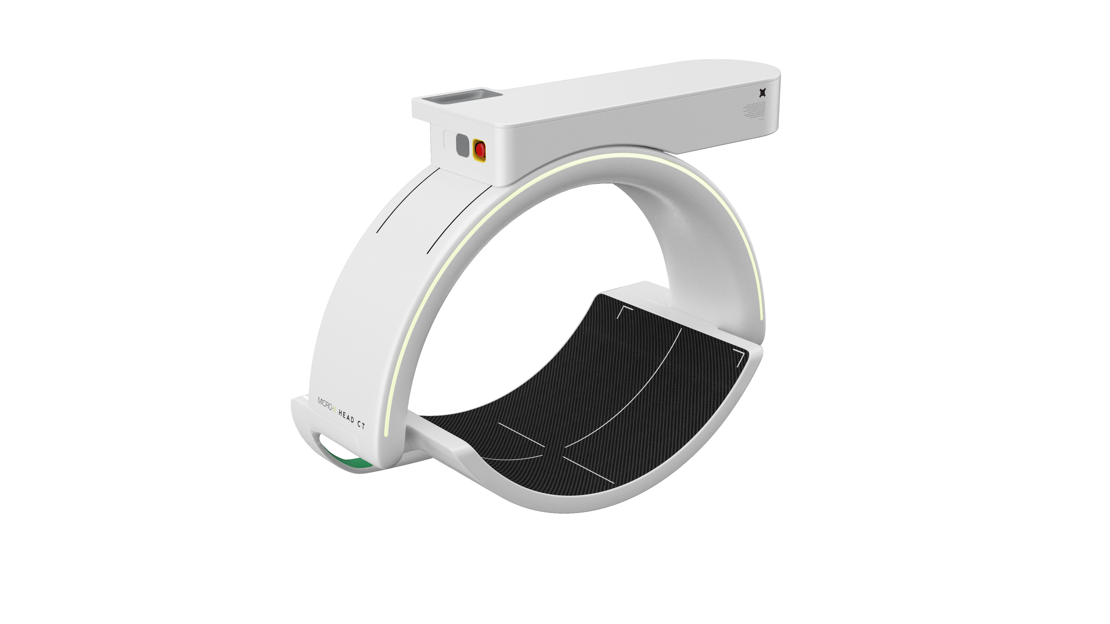 The Micro-X Head CT for stroke diagnosis includes a world-first curved detector
A World-First Achievement
To our knowledge, the Micro-X Head CT is the first CT scanner designed for clinical use to feature a curved detector. It’s a major technical milestone - not just for us, but for the future of portable medical imaging.
As an engineer, it’s been incredibly rewarding to push the boundaries of what’s possible in medical imaging. But what’s most meaningful is knowing this technology will have a real-world impact — transforming how and where people receive life-saving stroke care.
If you detect any issues, problems, or errors in this press release content, kindly contact [email protected] to notify us (it is important to note that this email is the authorized channel for such matters, sending multiple emails to multiple addresses does not necessarily help expedite your request). We will respond and rectify the situation in the next 8 hours.
Questions around this news
Why use a curved detector in a portable CT scanner?
The release says a curved detector gives the scanner wide angular coverage from a fixed gantry, avoiding heavy rotating components. Micro-X says this approach helps reduce the system’s size, weight, and power requirements, which are important for use in settings such as ambulances, rural clinics, and hospital emergency departments.
How was the curved detector developed?
Micro-X and FUJIFILM worked together on a custom detector, using FUJIFILM’s flexible scintillator substrate. The release describes overlapping two scintillators to form a curved array and pairing it with a rapid readout system. Micro-X later redesigned the detector, reporting a 72% weight reduction from the initial prototype.
What challenges did the team address in developing the imaging system?
The release says conventional reconstruction algorithms were not designed for curved detector geometry, which could cause inconsistencies and image artifacts. Micro-X worked with Johns Hopkins University on methods to address those issues. The system also uses Micro-X’s NEX technology to control X-ray sources without mechanical motion and capture multiple projections in rapid succession.
What is a scintillator and what does it do in an X-ray detector?
A scintillator is a material that emits light when struck by X-rays or other ionizing radiation. In many imaging systems, detector components capture that light and convert it into electrical signals, which can then be processed to form an image. Different scintillator materials and detector designs suit different imaging needs.
Earlier from Micro-X
- Cold Cathode X-ray Technology and its Advantages Over Conventional X-ray
- First look at the workflow for world-first mobile stroke diagnosis device
- Senior US Homeland Security leader visits Micro-X headquarters
- Checkpoints at Consumer Electronics Show 2024
- Micro-X joins the Veterans’ Employment Commitment
